
TLDR: The terms for these pregnancy tools may be used interchangeably, but they’re different: ultrasound is the imaging tool while sonogram is the image. Here’s everything you need to know about why ultrasounds are used, how they work, and how often you’ll experience them during pregnancy.
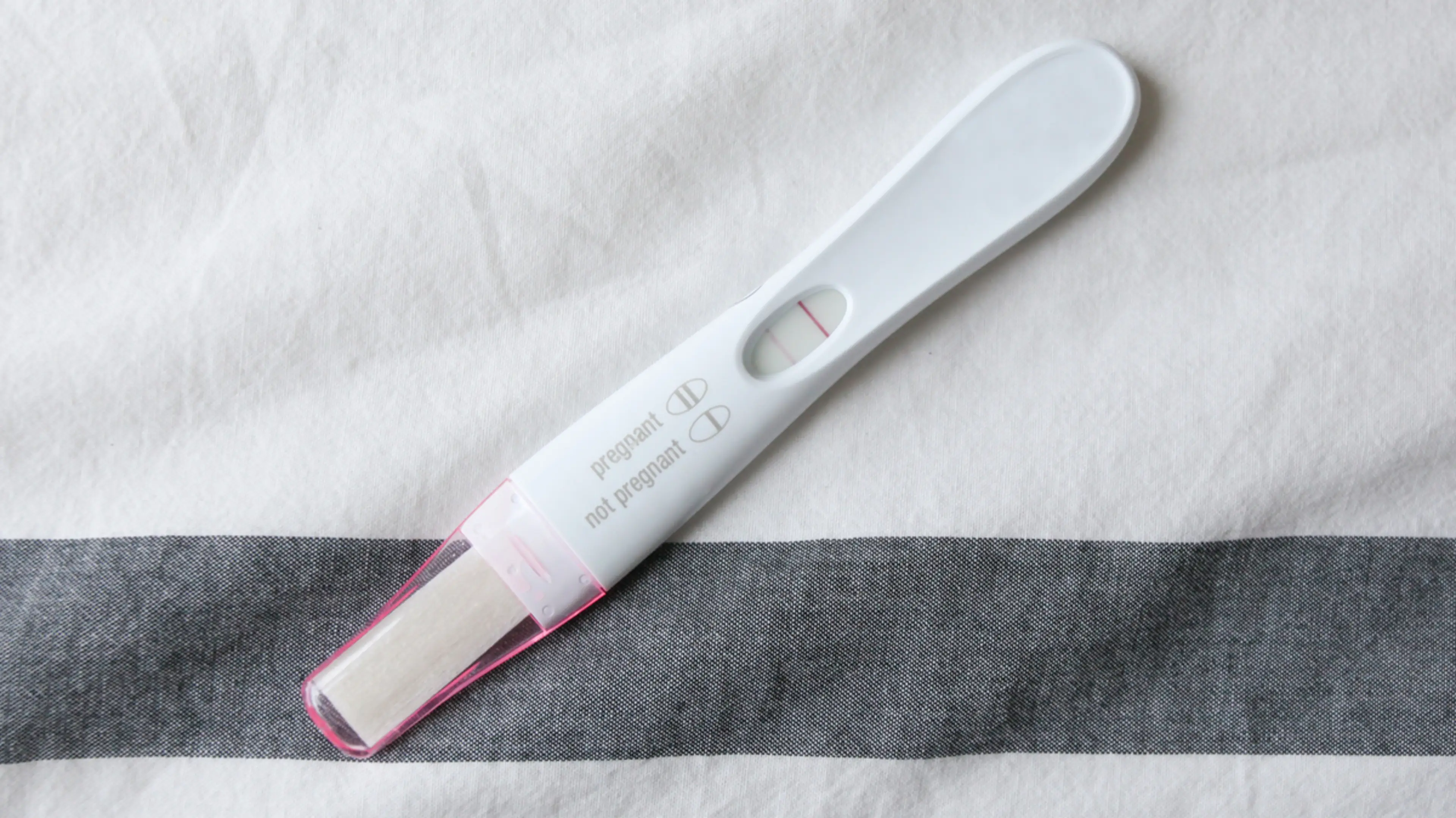
One of the hardest parts of the early pregnancy waiting game–other than the anticipation of watching those two pink lines to appear–is waiting to see that first image of your little one. You’ve probably heard the terms sonogram and ultrasound used interchangeably to describe how you’re able to see your baby in the womb. While they’re connected, they don’t actually mean the same thing.
Dr. Lauren Demosthenes, the senior medical director with Babyscripts1 , acknowledges that when you start your pregnancy journey, it’s easy to get them confused: “You may be told, ‘you are going to have an ultrasound,’ or ‘you are going to have a sonogram.’ I would not get hung up on the difference—the words are used interchangeably.”
While the terminology may not matter, understanding the different types of ultrasounds, how each is used during your pregnancy, and how often you should expect to experience them does.
What is an Ultrasound?
An ultrasound is a diagnostic machine that is used to view your baby during pregnancy. The first clinical use of an ultrasound was in 1956, but it didn’t start becoming widely used in pregnancy care until the 1980s. This exam is painless, non-invasive, and is used at different points in pregnancy.
An ultrasound uses sound waves to create pictures to see organs, blood vessels, soft tissue, and blood flow within the body. Unlike other diagnostic tools like x-rays, it doesn’t use radiation, so it’s a safer and more common exam used in pregnancy. With ultrasounds, doctors and techs can look for issues with the placenta and amniotic fluid, and see the baby’s position.
Your ob-gyn may also have a small, portable ultrasound that can be used for quick peeks but is usually not used for true diagnostics.
What is a Sonogram?
If the ultrasound is the camera that takes the images, the sonogram is the picture it produces. So, when you take that cute little black-and-white of your baby home, you are taking home a sonogram.
Sonograms can be hard to interpret, and you may be unsure about what you’re seeing, so don’t be afraid to ask the sonographer (another name for an ultrasound technician; these terms actually are interchangeable) or the doctor to explain. While only some of your ultrasound appointments automatically end with a photo (the big ones!), you can ask for a copy to take home after an ultrasound.
How does an Ultrasound Work?
Ultrasound technology4 is fascinating: An ultrasound consists of a computer, a monitor, and a transducer. A transducer is a handheld wand connected to the computer by a cable, which the doctor glides against your stomach during the exam. There are special crystals inside the transducer, which produce sound waves, and those pass through your skin into your body. Those waves bounce off your organs and tissue back to the transducer, creating an echo. The computer captures those echoes to produce the images we see that show the position, shape, and structure of organs and tissue on the screen. These images—sonograms—can be saved and printed.
What are the Types of Ultrasounds?
When we think of ultrasounds, we usually think of the type that pregnant people get on TV–the wand-on-the-stomach variety. But that’s just one form of ultrasound. If your doctor schedules an ultrasound, you can just ask them what type you’ll be getting, so you‘re not surprised when you get into the room. Here are the two different types of ultrasound you may experience.
Transabdominal Ultrasound
The type of ultrasound most people are familiar with is called a transabdominal ultrasound. Transabdominal ultrasounds can show the health of your baby as well as the health of your uterus, fallopian tubes, and ovaries.
For this ultrasound, the tech will put a special lubricating gel on your belly. The tech will then take the transducer and move it slowly from the side of your abdomen. This type of ultrasound isn’t invasive or painful, though there may be slight pressure from the transducer. The biggest complaint is usually that the gel is cold.
Tip: When you make your ultrasound appointment, make sure to ask whether or not you need a full bladder. Some scans require you to drink water before the ultrasound because a full bladder can help to produce clearer images.
Transvaginal Ultrasound
A transvaginal ultrasound uses the same technology, but the transducer is specially made to be inserted in the vagina. As you can imagine, this scan can be a little more uncomfortable than the transabdominal ultrasound. Transvaginal ultrasounds allow your doctor to see more detail in a smaller area. They are often used during fertility treatments like IVF as they can show the health of a woman’s reproductive organs.
When you get a transvaginal ultrasound, the sonographer will use lubricating gel on the transducer and insert it into your vagina. You may feel slight pressure or discomfort as they move the transducer around to get the images they need.
Sometimes in the same appointment, a transabdominal ultrasound will be used first to get the bigger picture, followed by a transvaginal ultrasound to see smaller details, or vice versa. The method your doctor chooses depends on what they need to see.
3D Ultrasounds
You’ve probably seen images from a 3D ultrasound, which is done in the same way as a traditional ultrasound (so there are no additional risks), but the image it produces is more detailed. You can usually get a better look at your baby in the womb, and the advantage of them from a medical perspective is that your doctor can also see any potential abnormalities better. While 3D ultrasounds aren’t standard, many offices do offer them—just ask your doctor or technician if you can expect one.
When and Why are Ultrasounds Used?
Ultrasounds are often used to monitor the health of the different reproductive organs in women. Because ultrasounds are generally safe, they’re used to monitor the health of babies in the womb and to try to detect reasons for infertility.
During Fertility
If you’ve been trying to get pregnant without success, your doctor may schedule you for an ultrasound to try to figure out what’s going on. When that appointment will be made depends on your age. If you’re under 35, it will likely be ordered once you’ve been trying for over a year. If you’re older than 35, they may recommend one after six months of trying. And if you’re 40 or older, you may go in for an ultrasound as soon as you tell your doctor you’re trying to conceive.
During an ultrasound to evaluate fertility3 , the sonographer will measure the lining of the uterus and ovaries as well as look for any abnormalities like fibroids or ovarian cysts. They will also be able to see the follicles on your ovaries to detect ovulation.
During Pregnancy
If your pregnancy is not high risk, you’ll most likely have two ultrasounds at two important times during your pregnancy.
The first ultrasound is taken early in the first trimester to confirm that you are pregnant. This scan may also be used to verify your due date. During this appointment, the sonographer will look for the baby’s heartbeat and will measure the baby from crown to rump. If you happen to be pregnant with more than one baby, this is when that big reveal will happen.
The second scan is usually during your second trimester between 18 and 22 weeks. This ultrasound is called an anatomy scan.
During this ultrasound, the sonographer will measure your baby from crown to rump again. They will also measure other areas like the head and around their middle to make sure everything is on track. They’ll be able to capture images of the heart, kidneys, brain, spine, and stomach.
Bonus: at this ultrasound, you can usually find out the sex of your baby.
Non-pregnancy Conditions
Ultrasounds have other uses besides examining growing babies and women’s reproductive organs. Ultrasounds are used to examine the overall health of organs and tissues in both men and women. An echocardiogram is used to look at the heart, while transabdominal ultrasounds can also be used to look at the liver, spleen, and bladder. There’s also an ultrasound that can scan the breasts. Conditions like bladder stones, enlarged or inflamed organs, and fatty liver disease can all be diagnosed through ultrasound.
Are Ultrasounds Safe?
Ultrasounds are widely considered to be safe2 during pregnancy for both the mother and the baby because there’s no radiation involved, and again, most types are non-invasive. (While the transvaginal ultrasound is invasive, it’s also considered safe.)
Getting an ultrasound can end up being an exciting milestone during your pregnancy since you spend so much time feeling your baby, but not seeing them. With this technology, you can learn more about your baby as they develop—and get a sneak preview of the little person you’re due to meet in just a few months.